Sandy Bay Campus, Chemistry Building, Level 2, Rooms 217, 254-256
Every person working in this facility has to have an induction before commencing work. Please watch this video and put your details in the form located on the flammable liquids cabinet in the laboratory (to your left when you enter the facility).
This facility houses:
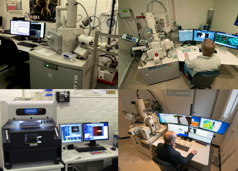
- JEOL JXA-8530F Plus field emission electron microprobe
- FEI MLA650 environmental scanning electron microscope
- Hitachi SU-70 field emission analytical scanning electron microscope
- digitised Olympus BX40F4 light optical microscope
- Cressington 208HR and BalTec SCD 050 sputter coaters
- Ladd 40000 and HHV Auto306 carbon evaporators
- Tousimis AutoSamdri 815 and Balzers CPD 030 critical point dryers
Electron Probe Microanalysis (EPMA) and Scanning Electron Microscopy (SEM) are closely related techniques for high-magnification imaging and spatially resolved chemical analysis of solid samples. Their focussed electron beam excites various secondary signals: Secondary electrons (SE) show surface morphology, backscattered electrons (BSE) local differences in mean atomic number. Crystal structure and orientation can be determined by electron backscatter diffraction (EBSD). Cathodoluminescence (CL) can be used to assess local variation in defect chemistry. X-rays are detected by wavelength and energy dispersive spectrometers (WDS, EDS) to measure variations in the local chemical composition. Whereas SEM is optimized for high-resolution microscopy, EPMA is mainly used for quantitative chemical analysis of micrometer-sized volumes, for which the samples have to be flat and polished. Non-conductive samples have to be coated (C, Au, Pt). Fresh biological specimens can be observed uncoated using the FEI in low vacuum or environmental mode.
Contact Dr Karsten Goemann or Dr Sandrin Feig for more information
This facility is part of Microscopy Australia

